February 20. 2008
Neo Caiel Servando was born at 7:30am..
He sleeps so peacefully... so quiet.... so cute! =)
He brought a different breeze in the family..
4months after.... he became cyanotic.. he woke up and cry in the wee hours in the morning but not that usual cry of babies.. it took hours of cry till dawn breaks. That's the time we seek the help of the experts. We went to various Doctors but then they had the same findings... My son has Congenital Heart Disease (Tetralogy of Fallot)
Tetralogy of Fallot (TOF) is a congenital heart defect which is classically understood to involve four anatomical abnormalities (although only three of them are always present). It is the most common cyanotic heart defect, and the most common cause of blue baby syndrome.
It was described in 1672 by Niels Stensen, in 1773 by Edward Sandifort, and in 1888 by the French physician Étienne-Louis Arthur Fallot, for whom it is named.
| Tetralogy of Fallot | |
|---|---|
| Classification and external resources | |
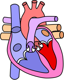
 Diagram of a healthy heart and one suffering from tetralogy of Fallot | |
| ICD-10 | Q21.3 |
| ICD-9 | 745.2 |
| OMIM | 187500 |
| DiseasesDB | 4660 |
| MedlinePlus | 001567 |
| eMedicine | emerg/575 |
| MeSH | D013771 |
Primary four malformations
"Tetralogy" denotes a four-part phenomenon in various fields, including literature, and the four parts the syndrome's name implies are its four signs. This is not to be confused with the similarly named teratology, a field of medicine concerned with abnormal development and congenital malformations, which thereby includes tetralogy of Fallot as part of its subject matter.
As such, by definition, tetralogy of Fallot involves exactly four heart malformations which present together:
| Condition | Description |
|---|---|
| A: Pulmonary stenosis | A narrowing of the right ventricular outflow tract and can occur at the pulmonary valve (valvular stenosis) or just below the pulmonary valve (infundibular stenosis). Infundibular pulmonic stenosis is mostly caused by overgrowth of the heart muscle wall (hypertrophy of the septoparietal trabeculae), however the events leading to the formation of the overriding aorta are also believed to be a cause. The pulmonic stenosis is the major cause of the malformations, with the other associated malformations acting as compensatory mechanisms to the pulmonic stenosis. The degree of stenosis varies between individuals with TOF, and is the primary determinant of symptoms and severity. This malformation is infrequently described as sub-pulmonary stenosis or subpulmonary obstruction. |
| B: Overriding aorta | An aortic valve with biventricular connection, that is, it is situated above the ventricular septal defect and connected to both the right and the left ventricle. The degree to which the aorta is attached to the right ventricle is referred to as its degree of "override." The aortic root can be displaced toward the front (anteriorly) or directly above the septal defect, but it is always abnormally located to the right of the root of the pulmonary artery. The degree of override is quite variable, with 5-95% of the valve being connected to the right ventricle. |
| C: ventricular septal defect (VSD) | A hole between the two bottom chambers (ventricles) of the heart. The defect is centered around the most superior aspect of the ventricular septum (the outlet septum), and in the majority of cases is single and large. In some cases thickening of the septum (septal hypertrophy) can narrow the margins of the defect. |
| D: Right ventricular hypertrophy | The right ventricle is more muscular than normal, causing a characteristic boot-shaped (coeur-en-sabot) appearance as seen by chest X-ray. Due to the misarrangement of the external ventricular septum, the right ventricular wall increases in size to deal with the increased obstruction to the right outflow tract. This feature is now generally agreed to be a secondary anomaly, as the level of hypertrophy generally increases with age. |
There is anatomic variation between the hearts of individuals with tetralogy of Fallot. Primarily, the degree of right ventricular outflow tract obstruction varies between patients and generally determines clinical symptoms and disease progression.
Additional anomalies
In addition, tetralogy of Fallot may present with other anatomical anomalies, including:- stenosis of the left pulmonary artery, in 40% of patients
- a bicuspid pulmonary valve, in 40% of patients
- right-sided aortic arch, in 25% of patients
- coronary artery anomalies, in 10% of patients
- a foramen ovale or atrial septal defect, in which case the syndrome is sometimes called a pentalogy of Fallot
- an atrioventricular septal defect
- partially or totally anomalous pulmonary venous return
- forked ribs and scoliosis
Diagnosis
The abnormal "coeur-en-sabot" (boot-like) appearance of a heart with tetralogy of Fallot is easily visible via chest x-ray, and before more sophisticated techniques became available, this was the definitive method of diagnosis. Congenital heart defects are now diagnosed with echocardiography, which is quick, involves no radiation, is very specific, and can be done prenatally.Treatment
Emergency management of tet spells
Prior to corrective surgery, children with tetralogy of Fallot may be prone to consequential acute hypoxia (tet spells), characterized by sudden cyanosis and syncope. These may be treated with beta-blockers such as propranolol, but acute episodes may require rapid intervention with morphine to reduce ventilatory drive and a vasopressor such as epinephrine, phenylephrine, or norepinephrine to increase blood pressure. Oxygen is effective in treating spells because it is a potent pulmonary vasodilator and systemic vasoconstrictor. This allows more blood flow to the lungs. There are also simple procedures such as squatting and the knee chest position which increases aortic wave reflection, increasing pressure on the left side of the heart, decreasing the right to left shunt thus decreasing the amount of deoxygenated blood entering the systemic circulation.[Palliative surgery
The condition was initially thought untreatable until surgeon Alfred Blalock, cardiologist Helen B. Taussig, and lab assistant Vivien Thomas at Johns Hopkins University developed a palliative surgical procedure, which involved forming an anastomosis between the subclavian artery and the pulmonary artery (See movie "Something the Lord Made"). It was actually Helen Taussig who convinced Alfred Blalock that the shunt was going to work. This redirected a large portion of the partially oxygenated blood leaving the heart for the body into the lungs, increasing flow through the pulmonary circuit, and greatly relieving symptoms in patients. The first Blalock-Thomas-Taussig shunt surgery was performed on 15-month old Eileen Saxon on November 29, 1944 with dramatic results.The Potts shunt and the Waterston-Cooley shunt are other shunt procedures which were developed for the same purpose. These are no longer used.
Currently, Blalock-Thomas-Taussig shunts are not normally performed on infants with TOF except for severe variants such as TOF with pulmonary atresia (pseudotruncus arteriosus).
Total surgical repair
The Blalock-Thomas-Taussig procedure, initially the only surgical treatment available for Tetralogy of Fallot, was palliative but not curative. The first total repair of Tetralogy of Fallot was done by a team led by C. Walton Lillehei at the University of Minnesota in 1954 on a 11-year-old boy. Successful total repair on infants has had success from 1981, with research indicating that it has a comparatively low mortality rate.Total repair of Tetralogy of Fallot initially carried a high mortality risk. This risk has gone down steadily over the years. Surgery is now often carried out in infants one year of age or younger with less than 5% perioperative mortality. The open-heart surgery is designed (1) to relieve the right ventricular outflow tract stenosis by careful resection of muscle and (2) to repair the VSD with a Gore-Tex patch or a homograft. Additional reparative or reconstructive surgery may be done on patients as required by their particular cardiac anatomy.
Prognosis
Untreated, tetralogy of Fallot rapidly results in progressive right ventricular hypertrophy due to the increased resistance on the right ventricle. This progresses to heart failure (dilated cardiomyopathy) which begins in the right heart and often leads to left heart failure. Actuarial survival for untreated tetralogy of Fallot is approximately 75% after the first year of life, 60% by four years, 30% by ten years, and 5% by forty years.Patients who have undergone total surgical repair of tetralogy of Fallot have improved hemodynamics and often have good to excellent cardiac function after the operation with some to no exercise intolerance (New York Heart Association Class I-II). Surgical success and long-term outcome greatly depends on the particular anatomy of the patient and the surgeon's skill and experience with this type of repair.
Ninety percent of patients with total repair as infants develop a progressively leaky pulmonary valve as the heart grows to its adult size but the valve does not. Patients also often have damage to the electrical system of the heart from surgical incisions, causing abnormalities as detected by EKG and/or arrhythmias.
Long-term follow up studies show that patients with total repair of TOF are at risk for sudden cardiac death and for heart failure. Therefore, lifetime follow-up care by an adult congenital cardiologist is recommended to monitor these risks and to recommend treatment, such as interventional procedures or re-operation, if it becomes necessary.
The use of antibiotics is no longer required by cardiologists and varies from case to case.
Now, my son is as active as a normal child at the age of 2. He still needs to undergo operation for him to survive. We are still looking for help to some organization who will help us financially for his operation. I know God is just there behind us and he knows what will happen next.


I was asked by a medical doctor Nicola Whitehill to purchase Dr Itua herbal medicine for Scleroderma as June is Scleroderma month. It took a lot to go through and remember that I have been through a lot! I have learned a lot about herbal medicines and my fight to have a healthy lifestyle! I have recommended Dr Itua herbal medicines to a lot of people so that they can see that the fight may be hard at times, but it is all worth it! You are worth the fight, so don't ever give up and continue to be your own advocate and you may need to switch doctors to get the best care for your body! Keep your head up and keep moving forward despite the obstacle that you may be on! God only gives you what He can handle, so put your faith, trust, and hope in Him and ask Him to show you the direction in which He wants you to go! By researching and finding what you are putting in or on your body can help tremendously and help you to use Food as Medicine to help heal your gut!
ReplyDeleteDr Itua has all types of herbal medicines to cure all kinds of disease such as Herpes,Diabetes,HPV,Copd,Als,Ms,HIV,Cancers,hepatitis,Parkinson,Infertility and other human disease and infections you may have been going through in your life Dr Itua will prepare you a permanent cure.
Dr Itua herbal center email contact: drituaherbalcenter@gmail.com. www.drituaherbalcenter.com